What Is Flow Cytometry?
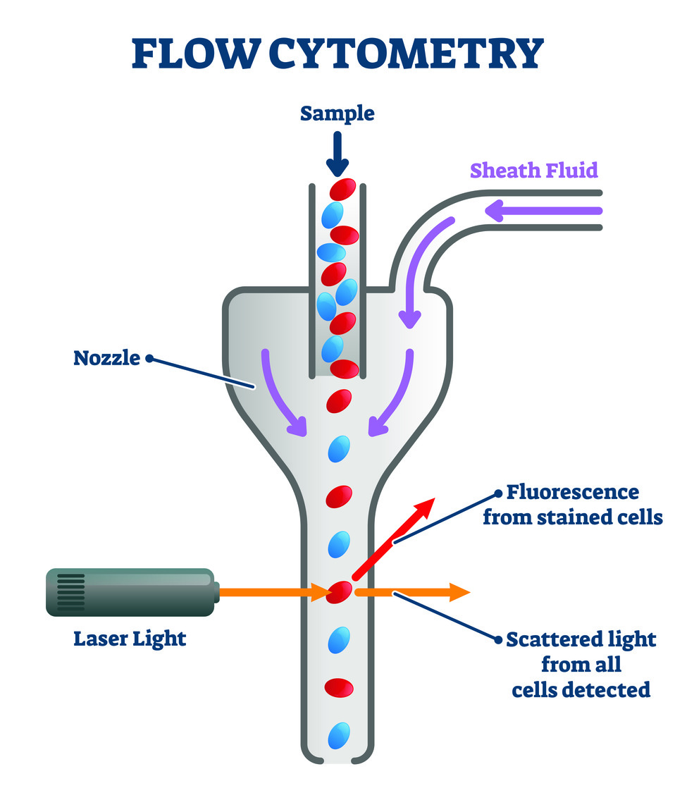
Flow cytometry is a method for counting and analyzing cells in a liquid suspension. Within a flow cytometer, lasers are a catalyst for creating quantifiable data based on the way sample components interact with light. A fluidics system enlists the aid of a sheath fluid, such as phosphate-buffered saline (PBS) to deliver the sample toward an interrogation point for analysis. The system utilizes hydrostatic pressure to guide the fluid, facilitating the laminar flow that causes cells to pass through the cytometer’s flow chamber in a single file line. As cells cross the beam(s) within, an optic system captures light scattering and/or fluorescence qualities, whereafter sophisticated software converts the data into voltage pulses, and generates graphical representations based on the collected information. The result is swift identification and organization of cells in a population.
From One, Many
The first commercially available flow cytometer was able to measure one or two parameters at a time. This early model entered the market in 1968, and the technology has since then evolved into modern flow cytometers that are routinely capable of analyzing as many as fifty cell traits simultaneously. Highly compatible with other forms of research, and able to drastically increase sample throughput, flow cytometry is often a core component in various other research methods. Three that work in conjunction include bead analysis, mass spectrometry, and microscopy. In fact, flow cytometry is so useful as a method of sorting, counting, and examining cells, that flow cytometers are common within more complex, multi-function laboratory equipment. Let us have a look at a few of its uses.
Immunophenotyping

The most widely used application of flow cytometry is immunophenotyping, and it is excellent for observing immune system response to autoimmune diseases and cancers. As part of the process, researchers stain cells with fluorochrome-conjugated antibodies. The cells, stained so, react to the interrogation point’s influence by giving off light of their own, and the results further serve to inform research efforts by revealing the presence of certain cell-surface markers (proteins) among cell populations. This is a basic look into immunophenotyping, but variations in application allow researchers to dive deep into the diverse mysteries of cell operation. Today, laboratories use immunophenotyping to aid in diagnoses, and in tracking the effectiveness of treatments for certain immunodeficiency disorders and leukemias.
Infectious Disease Monitoring

One discovered use of flow cytometry that led to swift acquisition of fluorescence flow cytometers in the 1980’s was in tracking HIV. HIV replicates on the surface of CD4+ T-cells, destroying them in the process. These cells have many functions that aid in fighting bodily invaders, including: instructing B-cells in which antibodies to produce, activating immune response toward cancerous cells, and in maximizing the effectiveness of bacteria-destroying agents. HIV whittles away at CD4+ T-cell population over time, eventually giving way to AIDS when cell count dips below a certain threshold. Almost four decades after these discoveries, modern flow cytometry continues to be a key component of HIV monitoring and research, as well as for other epidemics like tuberculosis and malaria.
Veterinary Medicine

As with research of the human body, flow cytometry plays a vital role in veterinary medicine. Researchers employ flow cytometry toward the same ends as the previously mentioned disciplines, diving into questions regarding immunology, oncology, and treatment progress. In dogs for example, researchers have used it to diagnose parasitic blood infections and in monitoring medication used for inflammatory and autoimmune issues. Just as for our own longevity, flow cytometry is a key component in the effort to extend the natural lifespan of our animal companions.
Something for Everyone
When it comes to counting, sorting, and analyzing cells, flow cytometry is an industry standard in immunology, but this is only one of many fields that harness the rich boon of today’s flow cytometers. The versatility in its applications make it a useful tool in fields ranging from forensic science; where it can be used to inform on things like criminal events and genealogy, to natural resources science; in which its uses include marking genetic differences between similar animal species, and monitoring the effects of dangerous chemicals on animal populations – such as in the event of oil spills.
Piedmont CMG’s Role

Bibliography:
Amiri, Storay. “Applications of Flow Cytometry in Veterinary Science.” News Medical, 22 Sep. 2022, www.news-medical.net/life-sciences/Applications-of-Flow-Cytometry-in-Veterinary-Science.aspx#2.
“Conjugated Primary Antibodies.” R&D Systems, www.rndsystems.com/products/conjugated-primary-antibodies.
“Fluidics of a Flow Cytometer.” ThermoFisher Scientific, www.thermofisher.com/us/en/home/life-science/cell-analysis/cell-analysis-learning-center/molecular-probes-school-of-fluorescence/flow-cytometry-basics/flow-cytometry-fundamentals/fluidics-flow-cytometer.html.
Fox, Amy et al. “Cyto-Feature Engineering: A Pipeline for Flow Cytometry Analysis to Uncover Immune Populations and Associations with Disease.” Nature Journal, 06 May 2020, www.nature.com/articles/s41598-020-64516-0.
“History of Flow Cytometry.” Beckman Coulter, www.beckman.com/resources/technologies/flow-cytometry/history.
Janossy, George and Shapiro, Howard. “Simplified Cytometry for Routine Monitoring of Infectious Diseases.” Wiley, 28 Jan. 2008, www.onlinelibrary.wiley.com/doi/full/10.1002/cyto.b.20405.
McKinnon, Katherine. “Flow Cytometry: An Overview.” National Library of Medicine, 21 Feb. 2018, www.ncbi.nlm.nih.gov/pmc/articles/PMC5939936/.
“T Helper Cell” Wikipedia, en.wikipedia.org/wiki/T_helper_cell.
Weir, Malcolm et al. “Flow Cytometry.” VCA Animal Hospitals, www.vcahospitals.com/know-your-pet/flow-cytometry.
Wetland and Aquatic Research Center. “Flow Cytometry Applied to the Animal Kingdom in Studies of Natural Resource Science.” United States Geological Survey, 26 May 2016, www.usgs.gov/centers/wetland-and-aquatic-research-center/science/flow-cytometry-applied-animal-kingdom-studies#overview.


